On February 12 , the international academic journal Cancer Research published an article online entitled “Latency Associated Nuclear Antigen of Kaposi's Sarcoma Associated Herpesvirus Promotes Angiogenesis through Targeting Notch Signaling Effector Hey1”by Prof. Ke Lan’s lab at Institut Pasteur of Shanghai, Chinese Academy of Sciences.
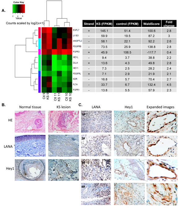
Notch signaling is implicated in the pathogenesis of Kaposi’s sarcoma (KS). KS is an angioproliferative neoplasm that originates from Kaposi’s sarcoma-associated herpesvirus (KSHV) infection. Previously, the researchers showed that the KSHV LANA protein can stabilize intracellular Notch in KSHV-infected tumor cells and promote cell proliferation. However, Notch signaling functions in pathological angiogenesis of KS remains largely unknown. Hey1, an essential downstream effector of the Notch signaling pathway, has been demonstrated to play a fundamental role in vascular development. In the present study, researchers perform whole transcriptome, paired-end sequencing on three patient-matched clinical KS specimens and their corresponding adjacent stroma samples, with an average depth of 42 million reads per sample. Dll4, Hey1 and HeyL display significant upregulation in KS. Further verification based on immunohistochemistry analysis demonstrates that Hey1 is highly expressed in KS lesions. Using the matrigel plug assay, down-regulation of Hey1 and gamma-secretase inhibitor (GSI) treatment causes dramatic reduction in the formation of new blood vessels in mice. Interestingly, LANA is responsible for the elevated level of Hey1 through inhibition of its degradation. More important, Hey1 stabilized by LANA promotes the neoplastic vasculature. The data suggest that hijacking of the pro-angiogenic property of Hey1 by LANA is an important strategy utilized by KSHV to achieve pathological angiogenesis.
The research was supervised by Prof. Ke Lan in collaboration with Prof. Hao Wen from Xinjiang Medical University and was completed by Postdoctoral fellow Wang Xing and other group members. The study was surpported by the Key project of Natural Science Foundation of China , the National Basic Research Program (973) and Sanofi - Aventis–SIBS scholarship.
The link for this article: http://cancerres.aacrjournals.org/content/early/2014/02/12/0008-5472.CAN-13-1467.long

Figure 1. LANA and Hey1 are highly expressed in pathological vascular endothelial cells in KS lesions. (A) Heat map representation of differentially regulated genes performed on RNA extracted from three KS lesions and their patient-matched normal tissues. The genes were ranked according to absolute values of log2 relative change. (B) LANA and Hey1 were highly specifically distributed in KS tumor rather than its adjacent stromal tissue on IHC analysis. Original magnification, ×5 in HE and ×40 in IHC. (C) IHC analysis of LANA and Hey1 co-localization in pathological vessels in KS lesions. Original magnification, ×40 in all examinations.

